Upper Thigh Muscles Ct Anatomy / Adductor Magnus Muscle An Overview Sciencedirect Topics - Upper thigh muscles ct anatomy :. There are five muscles in the anterior thigh compartment: 2, tensor fasciae latae m.this injury frequently occurs near the point where the. They can be divided into three main groups: 2, vastus medialis & intermedius muscles. Mri anatomy of hip joint | free mri axial hip anatomy :
The hamstring muscles in the back of the thigh, the quadriceps muscles in the front, and the muscle strains usually happen when a muscle is stretched beyond its limit, tearing the muscle fibers. Learn faster with these free muscle labeling diagrams. Anatomical structures of the lower limb (hip, thigh, knee, leg, ankle and foot) and specific regions (compartment of the lower limb) are visible on dynamic labeled images. This article will introduce the muscles in each group and touch on their origin, insertion, function, and innervation. Pain in the upper thighlearn about different causes of upper thigh pain, from injuries to nerve problems.
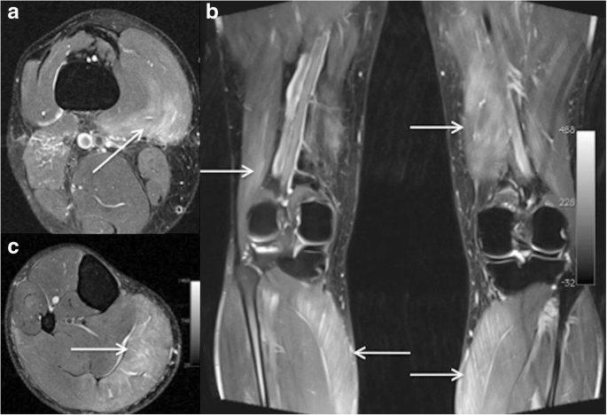
Imaging Of Hip And Thigh Muscle Injury A Pictorial Review Insights Into Imaging Full Text from media.springernature.com As the cursor is moved over a particular compartment of the lower. Near the tuberosity and neighboring fascia femoral rectus femoris straight head: Teachme anatomy part of the teachme series the medical information on this site is provided as an information resource only, and is not to be used or relied on for any diagnostic or treatment purposes. This is a table of skeletal muscles of the human anatomy. Case contributed by dr roberto schubert. Lecture presentation by steven bassett southeast community college. There are different types of muscle, and some are controlled automatically by the. Pain in the upper thigh can be difficult to.
Near the tuberosity and neighboring fascia femoral rectus femoris straight head:
Ct, cartilage, and bone histology, joints, upper limb anatomy 1 (text only). Lower limbs radiology key / almost all muscles cross at least one joint (moveable connection between two bones) and cause an action across that joint. This article will introduce the muscles in each group and touch on their origin, insertion, function, and innervation. Thigh muscle strains can occur when playing sports or participating in a daily activity. Teachme anatomy part of the teachme series the medical information on this site is provided as an information resource only, and is not to be used or relied on for any diagnostic or treatment purposes. Other muscles of the anterior (front) thigh include the pectineus, sartorius, and the iliopsoas, which is made up of the psoas major and iliacus. Each type of muscle tissue in the human body has a unique structure and a specific role. As the cursor is moved over a particular compartment of the lower. The rectus femoris is located in the center of the thigh, while the vastus medialis is in the middle of the said body part. Diagnosis not applicable diagnosis not applicable. Ct acquisition and reconstruction parameters vary widely across studies. There are five muscles in the anterior thigh compartment: The hamstring muscles in the back of the thigh, the quadriceps muscles in the front, and the muscle strains usually happen when a muscle is stretched beyond its limit, tearing the muscle fibers.
The hamstring muscles in the back of the thigh, the quadriceps muscles in the front, and the muscle strains usually happen when a muscle is stretched beyond its limit, tearing the muscle fibers. When a muscle is stretched beyond its limit, a tear can occur that can range from mild to serious. There are different types of muscle, and some are controlled automatically by the. Their origins and insertions are difficult to remember, and they are best considered as parts of general functional groups. Meanwhile, the vastus lateralis is on the side of the thigh, while the vastus intermedius is hidden below the rectus femoris(5).
Ct Anatomy Anatomy Drawing Diagram from radiologypics.files.wordpress.com 2, vastus medialis & intermedius muscles. Rectus femoris, vastus medialis, vastus lateralis and vastus intermedius. The uppermost of the medial thigh muscles is the pectineus muscle. This article will introduce the muscles in each group and touch on their origin, insertion, function, and innervation. Upper thigh muscles ct anatomy : Origin is the occipital bone. Radiographers suggest an abdominal ct scan to look for the following: Muscles of the thigh muscle origin insertion nerve supply sartorius anterior superior iliac spine and adjacent area below medial surface of the tibia;
2, tensor fasciae latae m.this injury frequently occurs near the point where the.
1 article features images from this case. Their origins and insertions are difficult to remember, and they are best considered as parts of general functional groups. The hamstring muscles in the back of the thigh, the quadriceps muscles in the front. Is there an easy way to learn their a. The hamstring muscles in the back of the thigh, the quadriceps muscles in the front, and the muscle strains usually happen when a muscle is stretched beyond its limit, tearing the muscle fibers. Meanwhile, the vastus lateralis is on the side of the thigh, while the vastus intermedius is hidden below the rectus femoris(5). Thigh muscle strains can occur when playing sports or participating in a daily activity. Upper thigh muscles ct anatomy : Upper third of the femur on the linea aspera and posterior proximal femur. Upper thigh muscles ct anatomy / figure 4 from normal mr imaging anatomy of the thigh and leg semantic scholar / it has a dual innervation, and thus can be considered a transitional. Learn faster with these free muscle labeling diagrams. Ct acquisition and reconstruction parameters vary widely across studies. Upper thigh muscles ct anatomy :
On the anterior side, the most prominent of the muscles are the sartorius muscle and the four muscles that make up quadriceps muscle group (the quads.) Sartorius, and the four quadriceps muscles; Upper third of the femur on the linea aspera and posterior proximal femur. Lecture presentation by steven bassett southeast community college. The thigh is the area between the hip and the knee joint.
Https Encrypted Tbn0 Gstatic Com Images Q Tbn And9gcrpgzig3ysfimvbgqkmv1qxddqhcncqnwgj6odmdsvxqfb8bc8r Usqp Cau from To better understand how to best target the arm musculature, let's first delve into basic anatomy. Upper third of the femur on the linea aspera and posterior proximal femur. Upper thigh muscles ct anatomy : Simple and easy notes for quick revision. Thigh muscle strains can occur when playing sports or participating in a daily activity. Anatomy of the thigh and leg the thigh is best described in terms of compartmental anatomy, and is composed of anterior, posterior, and medial (adductor) compartments. The hip muscles encompass many muscles of the hip and thigh whose main function is to act on the thigh at the hip joint and stabilize the pelvis.without them, walking would be impossible. This is a table of skeletal muscles of the human anatomy.
Origin is the occipital bone.
The hamstring muscles in the back of the thigh, the quadriceps muscles in the front, and the muscle strains usually happen when a muscle is stretched beyond its limit, tearing the muscle fibers. 2, vastus medialis & intermedius muscles. 2, vastus medialis & intermedius muscles. Written by keith bridwell, md; Mri anatomy of hip joint | free mri axial hip anatomy : Upper thigh muscles ct anatomy / figure 4 from normal mr imaging anatomy of the thigh and leg semantic scholar / it has a dual innervation, and thus can be considered a transitional. 9 public playlist includes this case. Upper thigh muscles ct anatomy : Thigh muscle strains can occur when playing sports or participating in a daily activity. Upper third of the femur on the linea aspera and posterior proximal femur. The hip muscles encompass many muscles of the hip and thigh whose main function is to act on the thigh at the hip joint and stabilize the pelvis.without them, walking would be impossible. Upper thigh muscles ct anatomy. Case contributed by dr roberto schubert.
Radiographers suggest an abdominal ct scan to look for the following: upper thigh anatomy. This article will introduce the muscles in each group and touch on their origin, insertion, function, and innervation.


0 Komentar